Header

Skip to main content

Slider

Peripheral Neuropathy & Amyloidosis

Neuropathy, also known as peripheral neuropathy, is a broad term that is used to describe damage to the nerves outside of the brain and spinal cord. There are over 100 types of peripheral neuropathy that can be classified into four categories, with each type having their own symptoms and prognosis. In this article, we’ll discuss the types of peripheral neuropathy and its connection to amyloidosis.


Symptoms

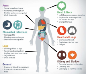
One of the challenges with neuropathy is the fact that symptoms can vary significantly based on what nerve is damaged. Additionally, symptoms can develop over the course of months to years (chronic neuropathy) or come on suddenly (acute neuropathy). Some of the most commonly seen symptoms are listed below:

  • Muscle weakness
  • Cramps
  • Muscle twitching
  • Loss of muscle and bone
  • Changes in skin, hair, or nails
  • Numbness
  • Loss of sensation or feeling in body parts
  • Loss of balance or other functions as a side effect of the loss of feeling in the legs, arms, or other body parts
  • Emotional disturbances
  • Sleep disruptions
  • Loss of pain or sensation that can put you at risk, such as not feeling an impending heart attack or limb pain
  • Inability to sweat properly, leading to heat intolerance
  • Loss of bladder control, leading to infection or incontinence
  • Dizziness, lightheadedness, or fainting because of a loss of control over blood pressure
  • Diarrhea, constipation, or incontinence related to nerve damage in the intestines or digestive tract
  • Trouble eating or swallowing
  • Life-threatening symptoms, such as difficulty breathing or irregular heartbeat


Types of Neuropathy

  1. Motor Neuropathy → Damage to the motor nerves control how you move.
  2. Sensory Neuropathy → Damage to sensory nerves control what you feel.
  3. Autonomic Nerve Neuropathy → Damage to autonomic nerves that control functions that are involuntary (ie. you do not consciously control).
  4. Combination Neuropathies → Damage to a mix of 2 or 3 of these other types of neuropathies. For example, damage to both sensory and motor nerves would result in sensory-motor neuropathy.


Amyloidosis

Peripheral Neuropathy is one of the hallmarks of amyloidosis, often seen in the transthyretin form of amyloidosis (ATTR). ATTR-PN, or transthyretin amyloid polyneuropathy, is a disease where the transthyretin protein becomes unstable and misfolds. This unstable protein (“amyloid”) then deposits in the nerve tissue, resulting in damage to these nerves. While amyloid deposits primarily in the peripheral nerves, it is not uncommon for amyloid deposition in the autonomic nerves as well. 

While peripheral neuropathy is most commonly associated with ATTR amyloidosis, it should be noted that peripheral neuropathy is also seen in 15-35% of patients with AL amyloidosis.

Most importantly, these are the most common and important signs and symptoms to be aware of, in order to diagnose ATTR amyloidosis.


Neurological Complications of ATTR Amyloidosis

Patients with ATTR amyloidosis are commonly faced with neurological complications. In this presentation, Dr. Chafic Karam from the University of Pennsylvania goes through four areas: an overview of the neurological systems, how amyloid damages the nerves, neurological signs of ATTR amyloidosis, and how to detect amyloid and diagnose ATTR amyloid neuropathy.


===========================================================

References:

https://my.clevelandclinic.org/health/diseases/14737-neuropathy

https://www.hopkinsmedicine.org/health/conditions-and-diseases/peripheral-neuropathy

https://www.mayoclinic.org/diseases-conditions/peripheral-neuropathy/symptoms-causes/syc-20352061

https://practicalneurology.com/articles/2021-july-aug/neuromuscular-amyloidosis

https://healthjade.net/familial-amyloidosis/


Proteinuria & Amyloidosis

According to the Cleveland Clinic, “Proteinuria is due to increased levels of protein in the urine.” Your kidneys filter waste products from your blood while retaining what your body needs — including proteins. However, some diseases and conditions allow proteins to pass through the filters of your kidneys, causing protein in the urine.


HOW DOES PROTEIN GET INTO URINE? (1)

Protein gets into the urine if the kidneys aren’t working properly. Normally, glomeruli, which are tiny loops of capillaries (blood vessels) in the kidneys, filter waste products and excess water from the blood.

Glomeruli pass these substances, but not larger proteins and blood cells, into the urine. If smaller proteins sneak through the glomeruli, tubules (long, thin, hollow tubes in the kidneys) recapture those proteins and keep them in the body.

However, if the glomeruli or tubules are damaged, if there is a problem with the reabsorption process of the proteins, or if there is an excessive protein load, the proteins will flow into the urine.


WHAT ARE THE SYMPTOMS OF PROTEINURIA? (2)

Often, someone with proteinuria doesn’t experience symptoms, especially if kidneys are just beginning to have problems. However, if proteinuria is advanced, symptoms can include:

  • More frequent urination
  • Shortness of breath
  • Tiredness
  • Nausea and vomiting
  • Swelling in the face, belly, feet or ankles
  • Lack of appetite
  • Muscle cramping at night
  • Puffiness around the eyes, especially in the morning
  • Foamy or bubbly urine

Conditions that can cause a temporary rise in the levels of protein in urine, but don’t necessarily indicate kidney damage, include:

  • Dehydration
  • Emotional stress
  • Exposure to extreme cold
  • Fever
  • Strenuous exercise

However, according to the Mayo Clinic (2), there are diseases and conditions that can cause persistently elevated levels of protein in urine, which might indicate kidney disease, such as:

TESTING FOR PROTEINURIA

The only way to know if you have protein in your urine, an established marker for chronic kidney disease, is to have a urine test.

“Integral to the process of evaluating for proteinuria is quantification of the total amount of protein spilling into the urine. The various methods to detect proteinuria include urine dipstick and sulfosalicyclic acid test (SSA); quantification methods include the ratio of albumin or protein to creatinine (UACR or UPCR) and the 24-hour urine protein collection.

The gold standard for quantification of proteinuria is the 24-hour urine collection. The test is performed by voiding upon waking and then collecting all urine on subsequent voids until the first void of the next day.“ (11)

In a retrospective study (5), researchers evaluated data from 265 patients with systemic AL amyloidosis who visited the Amyloidosis Center at Boston University Medical Center between July 1, 2018, and Jan. 1, 2020. This study examined the correlation between 24-hour urine testing and [urine protein-to-creatinine ratio] UPCR at various proteinuria levels in patients with AL amyloidosis. All patients underwent proteinuria measurement by 24-hour collection and UPCR in the same day. According to Andrea Havasi, MD, “In summary, although 24-hour urine collection is cumbersome, we continue to recommend it in patients with AL amyloidosis and kidney involvement.


CONCLUSION (12)

Amyloidosis can be a life-threatening disease because it can cause progressive organ damage and irreversible failure. Although it may affect any organ, one of the most frequently involved organs is the kidney, and clinically evident renal disease occurs in about 50-80% of cases. Typical manifestations of renal involvement are proteinuria, nephrotic syndrome (i.e., concomitant proteinuria, hypoalbuminemia, and peripheral edema), renal insufficiency, and end-stage renal disease (ESRD) requiring hemodialysis. All forms of systemic amyloidosis can lead to renal involvement. AL amyloidosis induces proteinuria and renal insufficiency in up to 73% and 50% of cases, respectively. ATTR amyloidosis typically does not involve the kidneys, but it can induce proteinuria and ESRD in some patients.


Therefore, when you have a patient with proteinuria, investigate why and don’t assume a benign origin. There are many serious causes, one of which may be amyloidosis.


Stay curious.


======== References  =========

  1. https://my.clevelandclinic.org/health/diseases/16428-proteinuria
  2. https://my.clevelandclinic.org/health/diseases/16428-proteinuria
  3. https://www.mayoclinic.org/symptoms/protein-in-urine/basics/causes/sym-20050656
  4. https://www.kidneyfund.org/kidney-disease/kidney-problems/protein-in-urine.html
  5. https://www.kidney.org/atoz/content/proteinuriawyska
  6. https://www.healio.com/news/nephrology/20220209/researchers-regard-24hour-proteinuria-collection-best-for-amyloid-light-chain-amyloidosis
  7. https://medlineplus.gov/lab-tests/albumin-blood-test/#:~:text=Albumin%20is%20a%20protein%20made,and%20enzymes%20throughout%20your%20body.
  8. https://www.kidney.org/content/kidney-failure-risk-factor-urine-albumin-to-creatinine-ration-uacr
  9. https://www.hopkinsmedicine.org/health/treatment-tests-and-therapies/24hour-urine-collection#:~:text=A%2024%2Dhour%20urine%20collection%20is%20a%20simple%20lab%20test,is%20returned%20to%20the%20lab
  10. https://www.kidneyfund.org/all-about-kidneys/tests-for-kidney-disease/urine-tests
  11. https://www.mdedge.com/clinicianreviews/article/210146/nephrology/proteinuria-and-albuminuria-whats-difference
  12. https://emedicine.medscape.com/article/238158-workup
  13. Talamo G, Mir Muhammad A, Pandey MK, Zhu J, Creer MH, Malysz J. Estimation of Daily Proteinuria in Patients with Amyloidosis by Using the Protein-To-Creatinine ratio in Random Urine Samples. Rare Tumors. 2015 Feb 18;7(1):5686. doi: 10.4081/rt.2015.5686. PMID: 25918613; PMCID: PMC4387359.

https://www.ncbi.nlm.nih.gov/pmc/articles/PMC4387359/#:~:text=AL%20amyloidosis%20induces%20proteinuria%20and,50%25%20of%20cases%2C%20respectively.&text=ATTR%20amyloidosis%20typically%20does%20not,and%20ESRD%20in%20some%20patients


Expert Insights: Amyloidosis – A Brief Clinical Overview

Dr. Sarah S. Lee, Assistant Professor, Division of Hematology, at the City of Hope, provides a brief yet comprehensive clinical overview of amyloidosis. In this video Dr. Lee discusses the breadth of amyloidosis, the wide range of symptom presentations, and which organs are typically involved. Focusing on AL (light chain) and TTR (transthyretin) types, she then goes through a diagnostic workup to arrive at a diagnosis, stressing the importance of typing once the presence of amyloid has been confirmed. Concluding her overview, Dr. Lee describes treatments available and how they impact patient prognosis and quality of life.


Cardiac Clues that Suggest Transthyretin Amyloidosis – Part I

In part 1 of a 2-part series, Dr. Keyur Shah, cardiologist from VCU Health’s cardiac amyloidosis care team, discusses the two most common types of transthyretin (TTR) amyloidosis: hereditary and wild-type. He details how ATTR cardiomyopathy amyloidosis presents and manifests itself to impair the heart. Dr. Shah lists clinical clues, “red flags,” and biomarkers which can raise suspicion of the presence of amyloidosis. Next he discusses insights that can be gained from echocardiograms, electrocardiograms, and cardiac MRIs and how they offer possible indicators of the disease presence. Once amyloidosis is suspected, definitive diagnosis testing is next.  See Part II: “Clinical Signs that Suggest Transthyretin Amyloidosis: Non-cardiac Clues” for more information.

Bicep Bunching & Amyloidosis


Often called “Popeye Deformity,” bicep bunching is visible when the patient flexes their arm, giving the appearance of Popeye-like arms. While it is the result of a torn tendon, it can be a leading indicator of more serious issues.


WHAT IS IT?

When the bicep tendon is ruptured, patients develop a bunching of the biceps upon flexion of the arm against gentle resistance. Tendon ruptures occur largely in the dominant arm of each patient, with one-quarter of patients developing ruptures in both arms. Interestingly, of those who had a rupture, 37.8% didn’t know it.


Below watch a video from The Lancet showing what bicep bunching looks like.


WHAT DOES IT POTENTIALLY INDICATE?

Two things.

1.  Bicep bunching may be a marker for ATTRwt. According to MedPage Today, spontaneous ruptures of the distal biceps tendon may be a marker of wild-type transthyretin (TTR) cardiac amyloidosis, a single-center study found. The presentation of a tendon rupture, an easily elicited diagnostic sign, in a patient with HFpEF should raise suspicion for wild-type TTR cardiac amyloidosis.

The picture below (Source: JAMA September 12, 2017 Volume 318, Number 10) offers examples of ruptured biceps tendon in two patients with biopsy-proven ATTRwt Cardiac Amyloidosis. ATTRwt indicates wild-type transthyretin amyloidosis. Patient 1 with prior rupture of the biceps tendon and bunching of the biceps with flexion. Patient 2 with acute rupture of the biceps tendon in the left arm; the tendon rupture occurred with trivial trauma, five years after Cardiac Amyloidosis diagnosis.

2.  ATTRwt may contribute to heart failure. Wild-type transthyretin amyloidosis (ATTRwt) is increasingly recognized as an important cause of heart failure with preserved ejection fraction (HFpEF).


WHY IS IT IMPORTANT?

Bicep bunching may be a marker of wild-type transthyretin (TTR) cardiac amyloidosis, potentially giving physicians an easy way to determine the underlying cause of heart failure with preserved ejection fraction (HFpEF) in some patients. Those who were aware, reported that the distal biceps tendon ruptured approximately five years prior to heart failure diagnosis, thus perhaps offering a leading insight.

In addition, early diagnosis of wild-type TTR cardiac amyloidosis (ATTRwt) is important because treatments are now available to slow, if not halt, disease progression. Unfortunately, the diagnosis of ATTRwt is often not considered in bicep bunching cases due to the perceived rarity of the disease.

“The clinical importance [of this study] is that the detection of a ruptured distal biceps tendon may be a clue for the diagnosis of wild-type TTR amyloidosis as the cause for heart failure. This diagnosis is often overlooked in clinical practice, so this relatively simple evaluation could increase detection of the disease,” said Stuart Katz, MD, of NYU Langone Health. “Enhanced detection could lead to better treatment.”


EXPERT INSIGHTS VIDEO ON MUSCULOSKELETAL MANIFESTATIONS

Dr. Shari Liberman, a hand and upper extremities surgeon from Houston Methodist Orthopedics & Sports Medicine, discussed six orthopedic manifestations and their pathology as it relates to systemic amyloidosis. Published studies, coupled with her experience, has led to a belief that these manifestations can offer important evidence of amyloidosis. She concludes with thoughts regarding an orthopedic differential and biopsy considerations for each of these manifestations.


Sources ———————————————————————————————————————
https://www.healthline.com/health/popeye-deformity
https://www.medpagetoday.com/cardiology/chf/67850
https://www.ncbi.nlm.nih.gov/pmc/articles/PMC5818850/
https://www.researchgate.net/publication/319657750_Association_Between_Ruptured_Distal_Biceps_Tendon_and_Wild-Type_Transthyretin_Cardiac_Amyloidosis
https://www.shoulderdoc.co.uk/article/721
https://www.thelancet.com/doi/story/10.1016/vid.2019.02.26.107679
https://www.youtube.com/watch?v=fHXu_0IZ3vU

Expert Insights: Why is amyloidosis so often misdiagnosed?

Dr. Angela Dispenzieri from the Mayo Clinic discusses why amyloidosis is often misdiagnosed. The complexity of the disease and commonality of symptoms are two of the reasons she examines. In addition, she offers guidance on appropriate diagnostic pathways.

Diagnosing Amyloidosis: A Two-Step Process

Amyloidosis can present in many types with the three most prevalent being light chain (AL) amyloidosis, hereditary variant transthyretin (ATTRv) amyloidosis, and wild type transthyretin (ATTRwt) amyloidosis. Being a rare disease, diagnosis can be particularly challenging, given that the general medical community is not well educated on the malady and symptoms are often associated with other more common ailments.

Successfully diagnosing the disease requires a two-step process before an appropriate treatment program can be determined and implemented for each patient.

  1. First, if amyloidosis is suspected, testing must be done to confirm the presence of amyloid.
  2. Second, once the presence of amyloid is confirmed, testing must then be done to identify and confirm the type of amyloidosis.

It is crucial that the second step, where the correct type of amyloidosis is identified, as the treatment regime can be different for each type. Here we share two different patient experiences which illustrate successful execution of the two-step diagnostic process.

Patient Case #1

The first case involved a 23-year old female. In 2017 she experienced an episode of coughing up blood, after which she looked in her throat with a flashlight and discovered a sizable lump. The patient met with a local ENT, who incorrectly diagnosed allergies, and prescribed over-the-counter medicine. With no improvement, she met with a second ENT. Testing was performed on the patient’s left oral pharynx utilizing a Congo red staining biopsy process which confirmed the presence of amyloid in the tissue. Additionally, mass spectrometry was performed which successfully differentiated the type of amyloidosis as being ALH (lambda light chain and delta heavy chain). Subsequently, she was referred to a hematologist who ordered a bone marrow biopsy and blood testing. The bone marrow biopsy summary notes read “….in conjunction with the concurrent finding of monoclonal lambda light chain restricted plasma cells in the marrow by flow cytometry, the findings are consistent with involvement of the marrow by a plasma cell neoplasm.”

Additionally, the blood testing confirmed elevated light chains as shown below.

Patient Case #2

The second case involved a man in his mid-fifties. He began experiencing disease symptoms approximately 6-7 years prior to being diagnosed in early 2019. He initially experienced gradually progressing numbness in his feet, legs, hands and forearms, as well as bilateral carpal tunnel syndrome. Soon after, he began experiencing symptoms of lightheadedness and fainting. Additionally, he started experiencing progressive gastro-intestinal issues such as acid reflux, chronic coughing, and frequent bouts of constipation and diarrhea. By 2018, his physical condition was rapidly deteriorating, including a total weight loss of approximately 80 pounds. During this extended period of time he was seen by a variety of physicians including internal medicine, neurology, endocrinology, gastroenterology, oncology, and cardiology, none of who were successful in arriving at a conclusive diagnosis. His list of maladies included cardiomyopathy, peripheral neuropathy, autonomic neuropathy, bilateral carpal tunnel syndrome, and gastroparesis, all which are classic symptoms of amyloidosis.

Finally, in early 2019 his condition was successfully diagnosed by an amyloidosis specialist. An echocardiogram was performed as well as a cardiac MRI (utilizing a gadolinium tracer) to identify amyloid fibrils and related damage in the heart tissue. These tests confirmed the presence of amyloid. A free light chain serum test was performed which ruled out AL amyloidosis, and Transthyretin DNA sequencing was performed to differentiate between the hereditary variant and wild-type of ATTR, which identified the T80A (legacy T60A) variant of transthyretin (ATTRv) amyloidosis. The two tests were successful in identifying the type of amyloidosis. The associated testing results are show below.

Echocardiogram Summary Notes

Associated Cardiac MRI Interpretation

DNA Sequencing Result


Once Diagnosed, Next is a Treatment Plan

Once the presence of amyloid is confirmed, and the type is identified, then it is time to treat the disease. In each of these patient cases the disease was diagnosed utilizing the two-step process to identify and confirm the type of amyloidosis. In both cases, successful treatment regimens were implemented which were effective in putting the disease into remission and/or halting disease progression.

Treatment options for amyloidosis have been vastly improved over the past several years. What was previously considered to be a foregone fatal disease can now be a manageable chronic disease. To ensure the best patient outcome, a timely diagnosis utilizing the two-step process, is essential.


Patient Insights: Seeking a second opinion

Our patient speakers at the Amyloidosis Speakers Bureau are powerful educators and offer compelling insights.

Have a listen to this brief clip from Mike sharing how it’s important for patients to self-advocate, particularly about their course of treatment. They are entitled to do research and if desired, seek a second medical opinion. How the physician reacts to the news can be telling.

Patient Insights: Best kept secret

Our patient speakers at the Amyloidosis Speakers Bureau are powerful educators and offer compelling insights.

Have a listen to this brief clip from Ozzie on his discovery of the ‘best kept secret’ as it pertains to diagnosing amyloidosis – carpal tunnel syndrome.

A Patient Guide for Understanding Amyloidosis

Amyloidosis is a multi-system disease, making diagnosis challenging. In this informative patient guide, the American Society of Nuclear Cardiology (ASNC) discusses common symptoms, types of amyloidosis, red flags to be aware of, diagnostic tests and available treatment options. 

CLICK HERE to read/download ASNC’s Guide for Understanding Amyloidosis


This website uses cookies

This site uses cookies to provide more personalized content, social media features, and ads, and to analyze our traffic. We might share information about your use of our site with our social media, advertising, and analytics partners who may combine it with other information that you’ve provided to them or that they’ve collected from your use of their services. We will never sell your information or share it with unaffiliated entities.

Newsletter Icon